
Craniotomy surgery
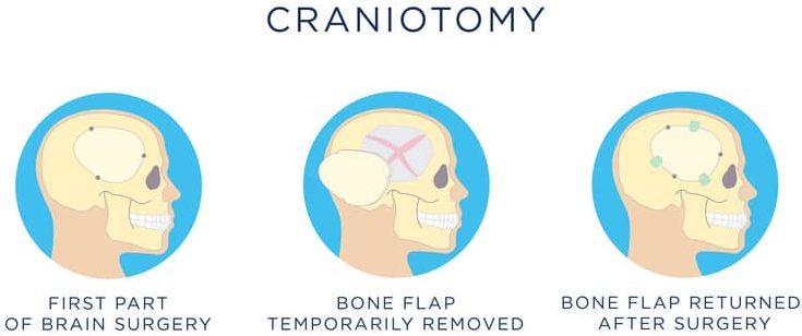
Brain surgery known as craniotomy necessitates the extraction of a section of the skull, or cranium, to allow access to the brain. After completion of the procedure, the bone is restored.
Typically, a craniotomy is performed for the purpose of eliminating brain tumors and addressing aneurysms.
The operation is carried out by a specialist in treating nervous system disorders. In this piece of writing, we will examine the different kinds of head surgeries, as well as the potential dangers and recuperation procedures involved.
Reasons for a craniotomy
A surgical procedure known as craniotomy is carried out to address various brain-related ailments.
- tumor
- aneurysm
- infection
- swelling (cerebral edema)
- bleeding inside the skull
- blood clot
- brain abscess
- skull fracture
- dura mater tear
- arteriovenous malformation
- arteriovenous fistula
- intracranial pressure
- epilepsy
The technique is additionally employed to insert implements for conditions related to locomotion, such as Parkinson’s disease.
Types of Craniotomy
Extended Bifrontal Craniotomy
The conventional method of reaching brain tumors located at the front involves an elongated incision in the skull, and is referred to as an extended bifrontal craniotomy. The rationale behind this approach is that it is less risky to extract additional bone than to handle the brain unnecessarily.
To perform an extended bifrontal craniotomy, the surgeon first creates a cut in the skin above the hairline, and then removes the bone that shapes the forehead and eye sockets. Following the surgical procedure, this bone is put back into its original position. This method enables the surgeon to operate in the area between the eyes, and directly behind them, while avoiding any unnecessary disturbance to the brain.
The method of extended bifrontal craniotomy is often employed to remove tumors that cannot be eliminated through minimally invasive means. Such tumors may be unsuitable for such approaches due to their anatomy, pathology, or surgical objectives.
The extended bifrontal craniotomy is utilized for the treatment of different kinds of tumors such as meningiomas, esthesioneuroblastomas, and malignant skull base tumors.
Posterior fossa craniotomy
The posterior fossa, situated in the base of the skull, is located in close proximity to the brainstem and cerebellum. The cerebellum is responsible for regulating balance and coordination.
In case there is a growth in the back area of the skull, it may cause compression on the cerebellum, brainstem, and spine.
To remove the tumor and relieve pressure, a surgical procedure called a posterior fossa craniotomy is performed by making an opening at the bottom of the skull.
Eyebrow Craniotomy
The “eyebrow” craniotomy, also known as supra-orbital craniotomy, is employed by doctors to eliminate brain tumors. To access tumors located in the frontal sections of the brain or close to the pituitary gland, which is positioned deeper in the brain behind the nose and eyes, a tiny cut is made in the eyebrow. This technique is utilized when the tumor is large or in proximity to crucial arteries or optic nerves, in lieu of endonasal endoscopic surgery.
the benefit of being a surgical technique that involves minimal incision. This procedure is known as supra-orbital craniotomy, as it involves the removal of a bone flap situated just above the eyebrow area.
- Less pain than open craniotomy
- Faster recovery than open craniotomy
- Minimal scarring
The procedure known as supra-orbital craniotomy can be utilized as a component of the therapy for certain medical conditions such as Rathke’s cleft cysts, tumors at the base of the skull, and certain types of pituitary tumors.
A craniotomy called “keyhole” is performed in the retro-sigmoid area.
The technique known as retro-sigmoid craniotomy, or “keyhole” craniotomy, is utilized as a less intrusive method to eliminate brain tumors. This approach is achieved by making a small cut behind the ear, enabling access to the cerebellum and brainstem, and the removal of tumors situated at the base of the skull. Medical professionals frequently make use of this technique to extract meningiomas, acoustic neuromas (vestibular schwannomas), skull base tumors, and metastatic brain tumors.
Advantages of “keyhole” craniotomy are diminished post-operative discomfort compared to open craniotomy, reduced scarring, and quicker recuperation.
Pteronial (frontotemporal) craniotomy
The point where the frontal, temporal, sphenoid, and parietal bones come together in the skull is known as the pterion. It is located in close proximity to the temple area on the side of the skull.
To gain access to different areas of the brain, a frontotemporal craniotomy, also known as a pteronial craniotomy, involves the removal of a section of the pterion. A surgical cut is made behind the hairline by the surgeon.
Orbitozygomatic Craniotomy
The orbitozygomatic craniotomy is an established method of accessing challenging tumors and aneurysms located at the base of the skull. The approach takes into account the fact that it is less risky to remove excess bone than to manipulate the brain without a valid reason.
Usually applied when less invasive methods are insufficient for addressing complicated lesions, orbitozygomatic craniotomy entails creating a scalp incision located behind the hairline and eliminating the bone that shapes the orbit and cheek. Subsequently, this bone is repositioned. This technique of temporarily removing the bone enables surgeons to access challenging, deeper areas of the brain with minimal impairment to it.
Orbitozygomatic craniotomy can be used as a treatment for brain tumors like craniopharyngiomas, pituitary tumors, and meningiomas.
Translabyrinthine Craniotomy
During a translabyrinthine craniotomy, the surgeon will make an incision at the back of your ear. They will then take out a section of the mastoid bone as well as the semicircular canals which play a crucial role in maintaining your balance.
A procedure is utilized to eliminate a vestibular schwannoma, which is alternatively known as an acoustic neuroma. This sort of growth is benign and develops on the nerve linking the inner ear and brain, resulting in problems with balance and hearing loss.
If the semicircular canals are removed, it can lead to a loss of hearing. Nonetheless, undergoing the surgery reduces the chance of damaging the facial nerve.
Stereotactic craniotomy
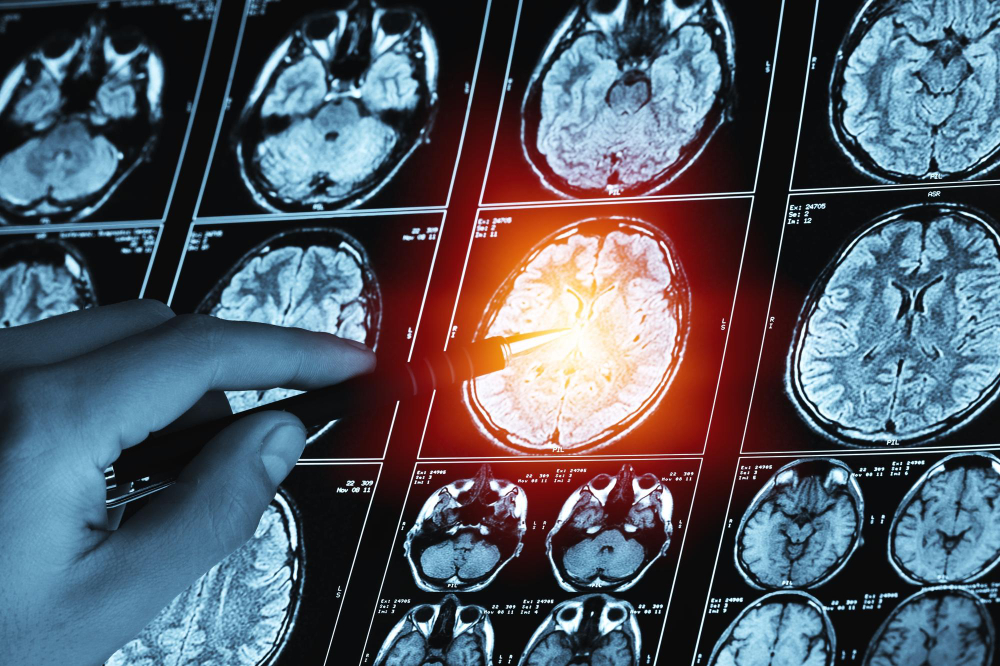
When an MRI or CT scan is utilized during a craniotomy, it is referred to as a stereotactic craniotomy.
Using imaging techniques, the surgeon produces 3D visual representations of your brain, which aid in the differentiation between normal and irregular tissue.
The use of stereotactic methods assists the surgeon in locating the ideal location for a scalp incision, making it simpler to create smaller incisions and carry out procedures that are less invasive.
Procedure for a craniotomy
The standard approach for performing a craniotomy consists of the subsequent stages.
- The hair on your scalp is shaved.
- You are given a general anaesthetic.
- In order to easily access the area of the brain injury, your head is positioned on a headrest that is either round or horseshoe-shaped. If it is necessary to limit movement of the head, a tool called a head pin fixing device may be used to hold the head in place.
- The neurosurgeon uses pre-surgery images to decide where to perform the craniotomy. To start the surgery, the scalp is cut open.
- The skull is punctured with a tool known as a perforator to create tiny openings, also known as burr holes.
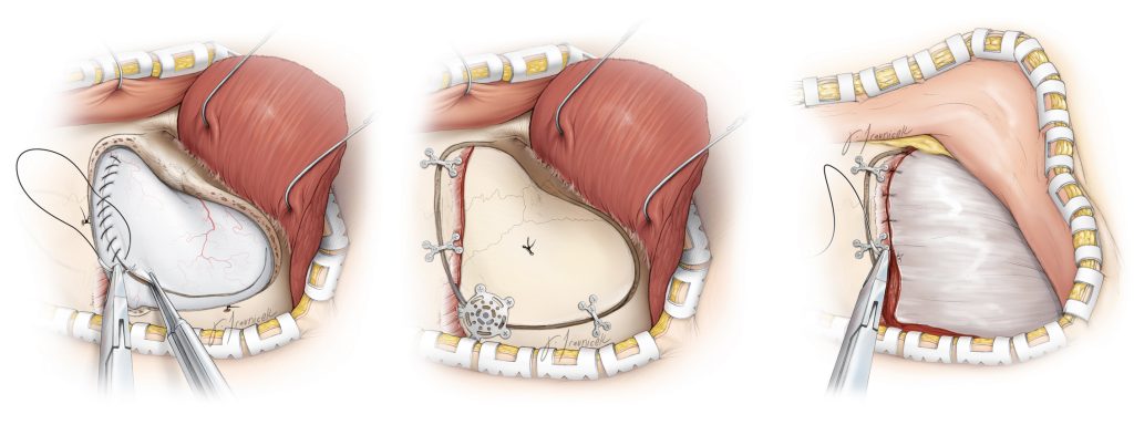
- To create a removable bone flap, a tool called a craniotome is employed to make incisions between two burr holes.
- Typically, the covering around the brain is lifted like a flap.
- The brain ailment undergoes a surgical procedure wherein repairs are made to the affected area such as fixing ruptured blood vessels or removing blood clots or tumors.
- Once the surgery is complete, the bone that was removed is put back in place, the muscle and skin are closed with sutures, and a drainage tube is inserted into the brain to eliminate any residual blood from the operation.
- It typically takes approximately two and a half hours to perform a craniotomy.
Recovery
After the surgery, a second meeting is scheduled within 10 to 14 days. The amount of time needed to recover ranges from 1 to 4 weeks, depending on the health condition being addressed and your overall well-being. To gradually raise your activity level, it is beneficial to walk. Be cautious not to overdo it, particularly if radiation or chemotherapy is still being administered. Inquire with the surgeon about when you might anticipate resuming work. It may take up to 8 weeks to fully recuperate.
Risks of the procedure
Complications are possible in any surgery, and brain surgery is particularly risky depending on which part of the brain is involved. If the region related to speech is operated on, it may result in speech difficulties. Other general possible complications include, but are not restricted to:
- Infection
- Bleeding
- Blood clots
- Pneumonia (infection of the lungs)
- Unstable blood pressure
- Seizures
- Muscle weakness
- Brain swelling
- when it escapes from where it is supposed to be located can cause various health issues. This may happen due to injury, surgery, a medical condition, or without any apparent cause. The symptoms can include headache, nausea, vomiting, sensitivity to light, and a clear or bloody discharge from the nose or ear. Treatment options may include observation, bedrest, medication, or surgery, depending on the severity and underlying cause of the leak.
- The utilization of general anesthesia possesses certain hazards.
Treatment in Türkiye:
The medical staff of surgical teams, doctors and consultants in Rehab Türk can provide the best treatment options and free consultations – by striving to keep abreast of the latest medical technologies and methods.