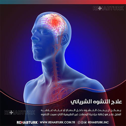
Cerebral arteriovenous malformation, diagnosis and treatment in Türkiye
Arteriovenous malformation (AVM) is an abnormal tangle of blood vessels in the brain or spine, some of which have no specific symptoms that pose minimal risks to an individual’s life or health, while others cause severe and devastating effects when bleeding occurs. Treatment options range from conservative observation to Surgery, based on the type of deformity, its symptoms and the location of its occurrence.
What is a cerebral arteriovenous malformation?
Blood flows from the heart through the large arteries to all regions of the body, and the arteries branch and shrink until they become capillaries, and there the exchange of fluids, oxygen and nutrients takes place, and waste picks up between the capillaries and body tissues in the capillary bed, but in the case of arteriovenous malformation, the arteries are directly connected to the veins without a capillary bed between them This creates a problem called high-pressure shunt, so the veins are unable to handle the blood pressure coming directly from the arteries, which leads to dilation and enlargement of the veins. Weak blood vessels can rupture and bleed, increasing the chances of developing an aneurysm.
Why do arteriovenous malformations occur in the brain?

Cerebral arteriovenous malformations are usually congenital, which means someone is born with the malformation. But they are usually not hereditary. It is possible that people do not inherit an AVM from their parents, and they probably will not pass one on to their children
Where do arteriovenous malformations occur in the brain?
Cerebral arteriovenous malformations can occur anywhere within or on the covering of the brain.
Do cerebral arteriovenous malformations change or grow?
Most AVMs do not grow or change much, although the vessels involved may dilate.
What are the symptoms of cerebral arteriovenous malformation?
Symptoms of AVM vary according to their type and location, but they do not show symptoms until they bleed. However, headaches and migraine-like attacks are general symptoms. Common signs of AVM in the brain include:
- Sudden onset of severe headache, vomiting and neck stiffness.
- seizures.
- Migraine-like headache.
- An abnormal ringing sound in the ear caused by the pulsation of blood through the AVM.
Common signs of a spinal arteriovenous malformation are:
- Sudden and severe back pain.
- Weakness in the legs or arms.
- paralysis.
What causes an AVM to bleed in the brain?
brain with arteriovenous malformation contains abnormal arteries and vessels. Therefore, the blood vessels that direct blood away from normal brain tissue are weakened.
These abnormal, weak blood vessels enlarge over time and may eventually burst from the high pressure of blood flow from the arteries.
What are the chances of bleeding in the brain?
The chance of a brain hemorrhage is 1% to 3% per year.
Does bleeding increase the chance of a second bleeding?
The risk of recurrent intracranial hemorrhage is slightly higher for the short time after the first hemorrhage. People ages 11 to 35 who have an AVM are at slightly higher risk of bleeding.
What can happen if a brain disease causes bleeding?
The risk of permanent brain damage is 20 to 30%. The probability of permanent brain damage is 20 to 30%. Every time blood leaks into the brain, normal brain tissue is damaged. This leads to a loss of normal function, which may be temporary or permanent.
Are there different types of brain arteriovenous malformations?
- Arteriovenous malformation: As we explained previously, it is of high pressure.
- Cavernoma: an abnormal group of enlarged capillaries with no important feeding arteries or veins and is of low pressure.
- Venous malformation: an abnormal group of enlarged veins that has low pressure and rarely bleeds and usually does not require treatment.
- Capillary telangiectasia: abnormal capillaries with enlarged areas, very low pressure, rarely bleed, and usually not treated.
- Dural arteriovenous fistula: abnormal connections between an artery and a vein in the tough membrane around the brain or spinal cord (dura mater).
An AVM forms anywhere arteries and veins are present. In the brain, AVMs can occur on the surface (the cortex) or in depth (in the thalamus, basal ganglia, or brainstem) and within the dura (the tough protective covering of the brain). Spinal arteriovenous malformations can occur on the surface (extramedullary) or inside the spinal cord (intramedullary) and are categorized into 4 types:
Type 1: This is a dural arteriovenous fistula and causes symptoms of venous hypertension.
Type 2: (also called the glomerular complex) is intramedullary.
Type 3: (also called juvenile) is an extensive arteriovenous malformation with abnormal intramedullary and extramedullary vessels.
Type 4: These are intradural arteriovenous fistulas.
What is the best treatment for a nerve fistula?
The best treatment is usually surgical endovascular resection of the abnormal connections that caused the fistula. This involves guiding small tubes (catheters) into blood vessels with X-ray guidance and blocking abnormal connections.
How is a cerebral arteriovenous malformation diagnosed?
If infection is suspected, the doctor takes a history of symptoms, current and past medical problems, current medications, and family history. A physical examination is also performed, and diagnostic tests are used to help determine the location, size, and type of AVM.
Among the diagnostic tests:
Computed tomography (CT) is an X-ray that is used to view the anatomical structures inside the brain. A newer technique called CT angiography injects contrast into the bloodstream to view the arteries of the brain. This type of test provides the best images of blood vessels by imaging the vessels and surrounding soft tissues.
Magnetic resonance imaging: (MRI) is a test that uses a magnetic field and radiofrequency waves to give a detailed view of the soft tissues of the brain. Angiography: This is an invasive procedure where a catheter is inserted into an artery and threaded through the blood vessels to the brain, contrast dye is injected once the catheter is in place in the bloodstream and x-ray images are taken.
What are the different types of treatment available?
Surgery, vascular therapy, and stereotactic radiosurgery alone are used in parallel to treat the AVM, but vascular embolization is used before surgery to reduce the size of the AVM and the risk of surgical bleeding.
Medical follow-up:
The doctor decides to monitor the patient if there has been no previous bleeding with anticonvulsants to prevent seizures and medications to lower blood pressure
Radiosurgery
Radiosurgery aims to direct precisely focused beams of radiation on abnormal vessels. Two main techniques are available: Leksell Gamma Knife and rainLab Novalis. The radiosurgery procedure takes several hours of preparation and one hour of radiation treatment. Then the patient goes home on the same day. The vessels are gradually closed and replaced with scar tissue after 6 months to 2 years.
- Advantage of radiosurgery:
- There is no surgical incision and the procedure is painless.
- Disadvantages of radiosurgery:
- Best with small AVMs and takes longer in terms of results.
Vascular embolization surgery
Using x-ray imaging, the doctor inserts a long, thin tube into the leg artery from the groin and threads it through the blood vessels to the brain region. Then the surgeon threads a catheter into one of the arteries feeding the AVM and injects an embolizing agent, such as a small particle or glue-like substance, to block the artery and reduce Blood flow in an arteriovenous malformation.
Craniotomy surgery
A surgical opening is made in the skull (using general anesthesia) and the brain is gently displaced at the site of the AVM. The AVM is then reduced and isolated from normal brain tissue using a variety of techniques such as lasers and electrocautery. However, it should be noted that the type of craniotomy performed depends on the size and location of the AVM
- Advantage of craniotomy surgery:
- The results are immediate if all the arteriovenous malformation is removed.
- Disadvantages of craniotomy surgery:
- Risk of bleeding, damage to nearby brain tissue, and stroke in other areas of the brain.
Treatment of cerebral arteriovenous malformation in Türkiye
Many patients come to Türkiye for the treatment of cerebral arteriovenous malformation. There are hospitals specializing in brain and nerve operations
REHABTÜRK HEALTHCARE PROVIDER NETWORK treatment services to patients in addition to transportation, accommodation and full trip coordination services.
Treatment of cerebral arteriovenous malformation in Türkiye requires at least two weeks for patients from abroad.
Patients receive intensive care after the operation to check their condition and satisfaction after the operation. In addition, our patient support team is available 24/7
The medical staff of surgical teams, doctors and consultants in REHABTÜRK can provide the best treatment options and free consultations – by striving to keep abreast of the latest medical technologies and methods.

